|
Nili Fossae in Natural Color and Across the Spectrum
- Click the image above for a larger view
- Full-Res JPEG (1448 x 1550) (298.6 kB)
- Full-Res TIFF (1448 x 1550) (6.7 MB)
Caption:
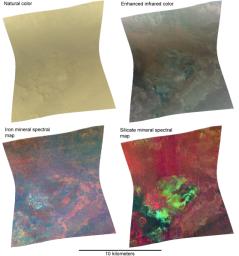
The Compact Reconnaissance Imaging Spectrometer for Mars (CRISM) took this image of the Nili Fossae region at 0643 UTC (2:43 a.m. EDT) on June 21, 2007, near 21.15 degrees north latitude, 74.24 degrees east longitude. CRISM's image was taken in 544 colors covering 0.36-3.92 micrometers, and shows features as small as 20 meters (66 feet) across. The region covered is just over 10 kilometers (6.2 miles) wide at its narrowest point, and is one of several dozen that CRISM has taken to map the minerals at candidate landing sites for the Mars Science Laboratory (MSL) mission, which will launch in 2010.
The Nili Fossae region is critical to understanding the history of water on Mars and whether water ever formed environments suitable for life, because the region is underlain by a layer of phyllosilicate (clay) minerals. This type of mineralogy formed where water was in contact with Mars' crustal rocks for very long periods, altering the silicates in volcanic rocks. In addition, phyllosilicates can encapsulate and preserve organic chemicals associated with life (if life was present). Its rocky record of an ancient wet environment makes Nili Fossae a top contender among the 30-plus landing sites being considered for MSL, whose objectives include measuring the chemistry preserved in an ancient wet environment.
This series of four different versions of the same 544-color image illustrates the mineral-mapping capability that comes from moving beyond the wavelength range of the human eye, and into infrared wavelengths where minerals leave distinct "fingerprints" in reflected sunlight. At upper left, more than three dozen of the distinct wavelengths measured by CRISM were combined to mimic how the human eye would see the image. The subtle shading comes from the Sun's position high in Mars' sky when the image was taken, creating few shadows. The bland, butterscotch color comes from the dust coating nearly all of the Martian surface to some degree. At upper right, three infrared wavelengths (2.53, 1.50 and 1.08 micrometers) replace the red, green and blue image planes. These wavelengths are less sensitive to dust, and begin to show the spectral variations in the underlying rocks.
The two bottom versions combine different wavelengths to show strength of absorption due to the different minerals that are present, providing indications of the minerals' presence and distribution. The lower left version combines measurements of the strength of iron mineral absorptions at 0.53, 0.86 and 1.0 microns in the red, green and blue image planes. Bluer areas have more pyroxene, a mineral found in volcanic basaltic rock, whereas reddish and especially orange areas have more oxidized iron minerals. The lower right version combines measurements of mineral absorptions at 1.0, 1.9 and 2.3 microns in the red, green and blue image planes. Redder areas are richer in pyroxene, and green and blue areas contain more phyllosilicate minerals. The combination of basaltic rocks and highly altered phyllosilicates in close proximity would allow MSL to make detailed measurements of rocks formed in two distinct environments.
Background Info:
The Compact Reconnaissance Imaging Spectrometer for Mars (CRISM) is one of six science instruments on NASA's Mars Reconnaissance Orbiter. Led by The Johns Hopkins University Applied Physics Laboratory, the CRISM team includes expertise from universities, government agencies and small businesses in the United States and abroad.
Cataloging Keywords:
| Name | Value | Additional Values |
|---|---|---|
| Target | Mars | |
| System | ||
| Target Type | Planet | |
| Mission | Mars Reconnaissance Orbiter (MRO) | Mars Science Laboratory (MSL) |
| Instrument Host | Mars Reconnaissance Orbiter | Curiosity Rover |
| Host Type | Orbiter | Rover |
| Instrument | Compact Reconnaissance Imaging Spectrometer for Mars (CRISM) | |
| Detector | ||
| Extra Keywords | Color, Dust, Infrared, Map, Shadow, Volcano, Water | |
| Acquisition Date | ||
| Release Date | 2007-09-11 | |
| Date in Caption | 2007-06-21 | |
| Image Credit | NASA/JPL/JHUAPL | |
| Source | photojournal.jpl.nasa.gov/catalog/PIA10073 | |
| Identifier | PIA10073 | |
